nalco group
bone, muscle & joint pain physio
BOOK NOW / WHATSAPP ABOUT YOUR PAIN OR INJURY
- NOVENA 10 Sinaran Drive, Novena Medical Center #10-09, Singapore 307506
- TAMPINES 9 Tampines Grande #01-20 Singapore 528735
- SERANGOON 265 Serangoon Central Drive #04-269 Singapore 550265
Home > Blog > Physiotherapy & Hand Therapy > Conditions > Elbow Pain > Radial Head Fracture Hand Physiotherapy
Radial Head Fracture Hand Physiotherapy

Dislocations and fractures of the radial head are among the most common elbow injuries in adults.
One in every six elbow fractures in hospital emergency departments involves damage to the radial head (that's close to 17% occurrence rate).
Radial head fracture injuries are typically characterized by:
- Pain
- Tenderness
- Bruising
- Swelling
- Loss of strength and
- Deformity in the elbow, forearm, and wrist
A radial head fracture may affect only the radial head and lateral side of the elbow. Or it may involve a complex injury pattern that affects other parts of the upper extremity, including the humerus, forearm, hands, and wrists.
Radial head fractures will require proper and prompt treatment to retain healthy joint function and prevent permanent disability.
Failing to do so can lead to
- elbow nerve
damage
- trauma-induced arthritis
- and similar complications
Conventional treatments include both open and closed methods and require hand therapy and elbow physiotherapy for complete recovery.
In this blog, we take an in-depth look at fractures of the radial head and what causes them. We also discuss the common symptoms and other injuries associated with radial fractures and explore the treatment options you should know about.
What Are the Fractures of the Radial Head?

The radial head is an articular surface near the proximal end or the central part of the elbow joint.
Basically, it is the crown of the radius, which, along with the humerus, constitute the two main bones in your forearm. The radius runs from the elbow joint to the outer side of the wrist, ending just where the thumb is located.
Any crack that develops in the radius right beneath the elbow is referred to as a fracture of the radial head.
Radial head fractures are usually classified into three main types, depending on the distance by which the bones have been displaced. The extent of displacement dictates the treatment plan and types of physical therapy required for recovery.
This occurs when the radius is displaced by less than 2 mm.
There might be just a split in the bone, and it may not have moved from its normal position at all. Type I fractures can be quite hard to diagnose initially as small cracks may not be visible on x-ray images. They generally become more prominent and show up on scans performed 2-3 weeks after the injury.
During this time, people usually experience pain in the elbow movement.
Performing any strenuous activities with a type I radial fracture can shift the bones significantly and worsen the condition.
Type II Fracture
If the radius is displaced by more than 2 mm, it is referred to as type II fracture.
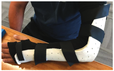
Type II fractures generally affect a large part of the bone and may hinder specific movements, such as forearm and wrist rotation. Patients may recover after wearing a sling or brace for 1 to 2 weeks, followed by mobility exercises.
However, depending on the damage caused, minor surgery may be required to remove small pieces of the broken bone, especially if they obstruct joint function.
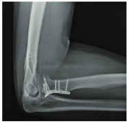
Where a large bone fragment has moved out of its regular place, a metal plate and screws may be necessary to join it back.
Type III Fracture
Type III fractures are the most severe kind of radial head injuries.
They produce multiple bone splinters that can make bone restoration almost impossible. In most cases, the damage extends to the ligaments, nerves, and other structures surrounding the elbow joint.
Type III fractures typically call for major surgery.
The surgeon may either remove the broken fragments altogether or, if possible, try to fix it by putting them back together. If the removal of the radial head is necessary, an artificial unit may be installed in its place.
This is a permanent treatment and can restore a degree of movement, ensuring long-term joint operation.
What Causes Fractures of the Radial Head?
In most
cases, there is a history of falling on an outstretched hand.

In senior people, radial head fractures may be caused by certain orthopedic conditions, such as rheumatoid arthritis and osteoporosis.
In younger patients presenting with radial head fractures, the injury usually stems from direct trauma to the joint. For example, athletic activities and motor vehicle accidents can result in damage to the radius and the radial head.
Symptoms of Radial Head Fractures
The most common symptoms of this type of elbow injury include:
- Joint ache
- Stiffness
- Numbness or tingling sensation
- Skin discoloration and swelling on the outer side of the elbow
- Intense pain while straightening or flexing the elbow
- Difficulty or inability to rotate the forearm (e.g., turning hand from palm down to palm up and vice versa)
People who fracture their radial head typically experience elbow pain and swelling soon after the fall or injury. If the bones are displaced, there may be an obvious deformity with the pointy edge of the elbow sticking out at an odd angle.
Individuals who develop type I radial head fractures may only experience minor discomfort at first.
However, with the passage of time, it can progress into more severe symptoms, including
- limited joint movement
- bruising and
- tenderness
Other Conditions Similar to Radial Head Fractures
There are hardly any conditions that are similar to a fractured radial head.
It’s a distinct type of injury that can be diagnosed with radiography tests or a clinical examination that assesses the biomechanics of the upper extremity.
However, individuals suffering from a radial head fracture tend to develop other elbow conditions or nerve conditions with this injury. This particularly happens when the fracture goes undetected due to minor bone displacement.
The conditions that can develop following the fractures of the radial head include:
- Posterior interosseous neuropathy
- Radiocapitellar arthrosis (posttraumatic joint arthritis)
- Annular ligament disruption
- Lateral ulnar collateral ligament (LUCL) tear
- Medial (ulnar) collateral ligament (MCL) strain/ rupture
Who Is at Risk of Developing Fractures of the Radial Head?
Everyone is at risk of fracturing their radial head, especially when they try to break a fall with their hands. Of course, it’s only an instinctive motion. But it shifts your entire weight onto the elbow and forearm muscles.
The impact of the fall can easily force the radius to move farther back into the joint, dislocating the radial head in the process.
Men and women above 40 years of age are also prone to fracturing the radial head. This is mostly due to the fact that bones naturally become weak and fragile with age. Therefore, even a seemingly minor strain on the forearm can increase their chances of experiencing radial head dislocation.
Athletes and other people who frequently partake in sports activities are also at a high risk of fracturing their radial head.
Similarly, construction workers and people with known medical conditions, such as osteochondritis and Panner’s disease, are also susceptible to radial head fractures.
Treatment for Fractures of the Radial Head
Radial head fractures are serious injuries that can have life-long consequences if not treated on time. If you think you may have fractured your radial head, seek medical help at your local hospital or emergency department immediately.
Individuals with a fractured or dislocated radial head are generally advised not to perform activities with the affected arm.
Until your injury is assessed by a medical practitioner, it’s better to avoid daily activities, such as hot showers, which can increase the rate of blood flow in the body, which in turn, will increase pain and swelling.
Your doctor may take an x-ray scan to
identify the degree of bone damage and displacement. Initial management
focuses on stabilizing the fracture so that the bones can set and heal
properly.
Whether your arm is placed in a cast directly or you need
to undergo surgery to set the bones in their regular position,
our senior physiotherapists and hand therapists will definitely be needed to restore joint function and
strength before you can resume full activities.
How Physiotherapy Can Help People with a Fractured Radial Head

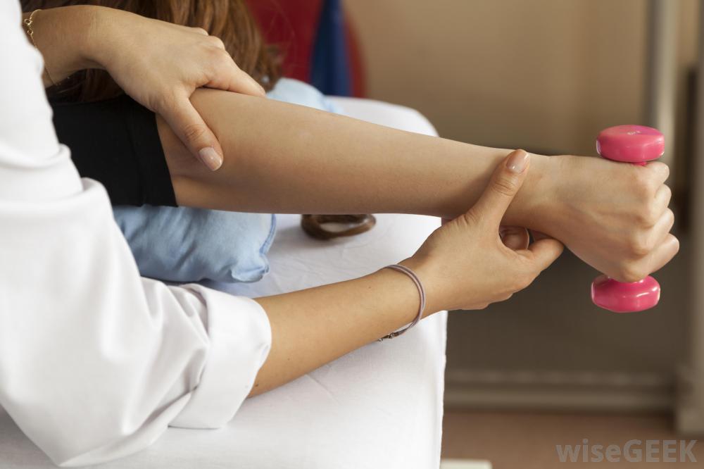
The main goal of physiotherapy following a radial head fracture or dislocation is to improve the range of motion of the affected arm. It also helps reduce the pain and swelling and is especially useful for improving recovery after surgery.
After initial treatment at a hospital, individuals can come to us for physical therapy for healing a fractured radial head.
Our master-level physiotherapists perform a thorough examination to determine which plan and mobility exercises will be best suited to each individual.
Physiotherapy for radial head fractures involves a progressive series of exercises. They are specially designed to promote natural bone restoration and gradually improve the range of motion of your injured arm. At times, other therapies might also be used in combination to ensure efficient and effective recovery.

Some of the therapeutic modalities we commonly use for fractured radial head rehab include:
- Joint mobilization
- Electrotherapy such as ultrasound therapy
- Heat therapy such as paraffin wax bath and heat packs
- Acupuncture
- Taping
- Soft tissue massage
Fractures of the radial head usually heal without complications within 6 to 8 weeks after the bone stabilization. Some individuals are able to resume lighter activities within 2 to 3 weeks as well.
The key is to seek medical help immediately and comply with the physical therapy plan so you can quickly get back to doing what you love.
